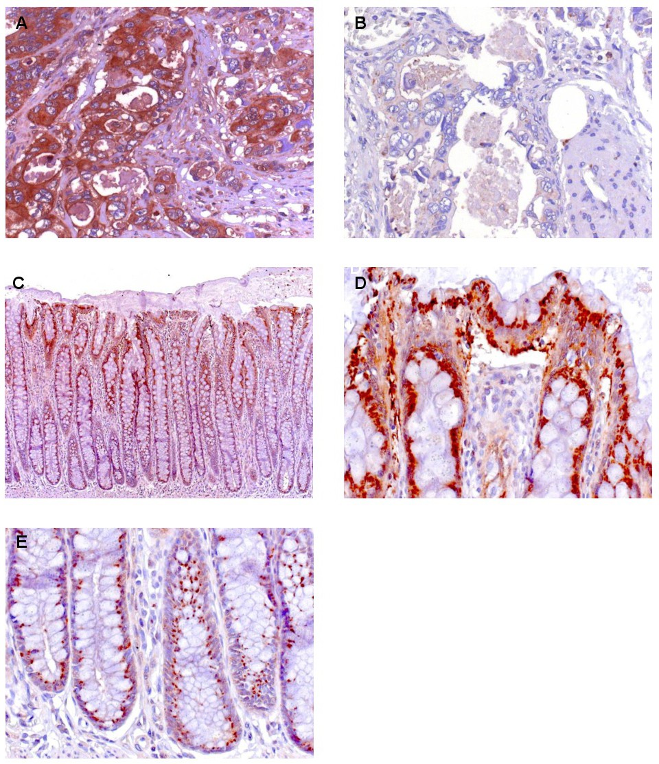
Fig. 1. MLK4 staining in human colorectal carcinomas (CRC) and normal colonic mucosa. Representative histological appearance of MLK4 staining with predominant cytoplasmic staining pattern (score 1 - A) or negative staining (score 0 - B) in different tumor specimens. Positive pattern of cytoplasmic staining of MLK4 in epithelial cell of normal colonic mucosa visible along the whole length of the crypts (C). Particularly high staining intensity in the apical regions of the crypts (D) and granular staining patter of MLK4 in cells of the crypt basis (E). Magnifications: × 100 (C) and × 400 (A, B, D, E).